| Electric Cell-Substrate Impedance Sensing |
| Electric
Cell-Substrate
Impedance
Sensing
or short ECIS has first been described in 1984 by Ivar
Giaever and Charles
R. Keese (Rensselaer Polytechnic Institute, Troy, NY; Applied
BioPhysics Inc., Troy NY). The basic principle of the technique is
sketched below.
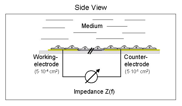
Thin
gold-films (100 nm) are deposited and layouted on the bottom of ordinary
cell culture substrates and the cells are grown on the surface of these
electrodes. The gold-electrodes are prepared thin enough to be transparent
so that the cells can be inspected by phase contrast microscopy. One of
the two electrodes, that are used in the measurement, is named the working
electrode and it is much smaller than the so-called
counter electrode.
The medium that overlays the cell layer is an ionic conductor and thereby
closes the electrical circuit. The principle of ECIS is to measure the
electrical
impedance of this system as a function of AC frequency.
The
measured impedance depends on the three dimensional shape of the cell body
and and reports on changes in cell shape upon any kind of experimental
challenge.
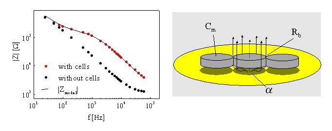
On the left side of the following figure you can see the electrical impedance as a function of frequency for a cell-free gold electrode (black circles) compared to the impedance of the same electrode when it is covered with a confluent layer of epithelial cells (red circles).
The presence of the cells on the electrode surface causes the impedance to increase significantly, but these increases are strongly dependent on the frequency of the applied electric field. The surface-attached cells behave essentially like insulating particles, that block the current flow from the electrode and thereby increase the electrode impedance. The impedance raw data of cell covered electrodes can be analyzed in terms of cell morphology by using a theoretical model that is sketched on the right hand side. Dependent on the frequency of the applied electric field a fraction of the current can either flow through the cells (transcellular) or is forced to flow around the cellular bodies (paracellular). The amount of transcellular current is dependent on the capacitance of the plasma membrane whereas the paracellular current is limited by the distance between cell and substrate as well as the resistance of the intercellular cleft. These three contributions can be distinguished and separated from each other mathematically. The analysis provides three parameters that describe the cell layer: 1.
Resistance between adjacent cells Rb
as a measure for the tightness of cell-cell-contacts.
We apply this technique as a whole-cell biosensor for a wide variety of problems and develop modifications that open up new applications. One aspect is the defined manipulation of the substrat anchored cells by means of invasive electric fields for electroporation or introduction of lesions. |
| Home | Group Members | Publications |